Split Dynamic MRI represents a breakthrough in medical imaging, utilizing high temporal and spatial resolution to revolutionize tumor diagnosis. This technique, by merging rapid sequence acquisition with detailed imagery, offers unparalleled precision in distinguishing between malignant and benign lesions. By providing clearer, more detailed images, Split Dynamic MRI aids clinicians in making more accurate diagnoses and treatment plans, particularly in complex cancer cases.
Key benefits include faster diagnosis times, reduced need for repeat scans, and the potential to identify cancers earlier in their development. This not only enhances patient care but also streamlines the diagnostic process in oncology.
We have thoroughly investigated the clinical utility of the Split Dynamic MRI data. Our findings indicate that high temporal resolution combined with dynamic T1 -weighted and R2* assessment provides a high diagnostic accuracy when differentiating malignant from benign breast masses. Our findings also indicate that the additional kinetic information, combined with BI-RADS interpretations from high spatial resolution image series, improves the diagnostic accuracy in the assessments of breast masses compared to BI-RADS interpretations alone.
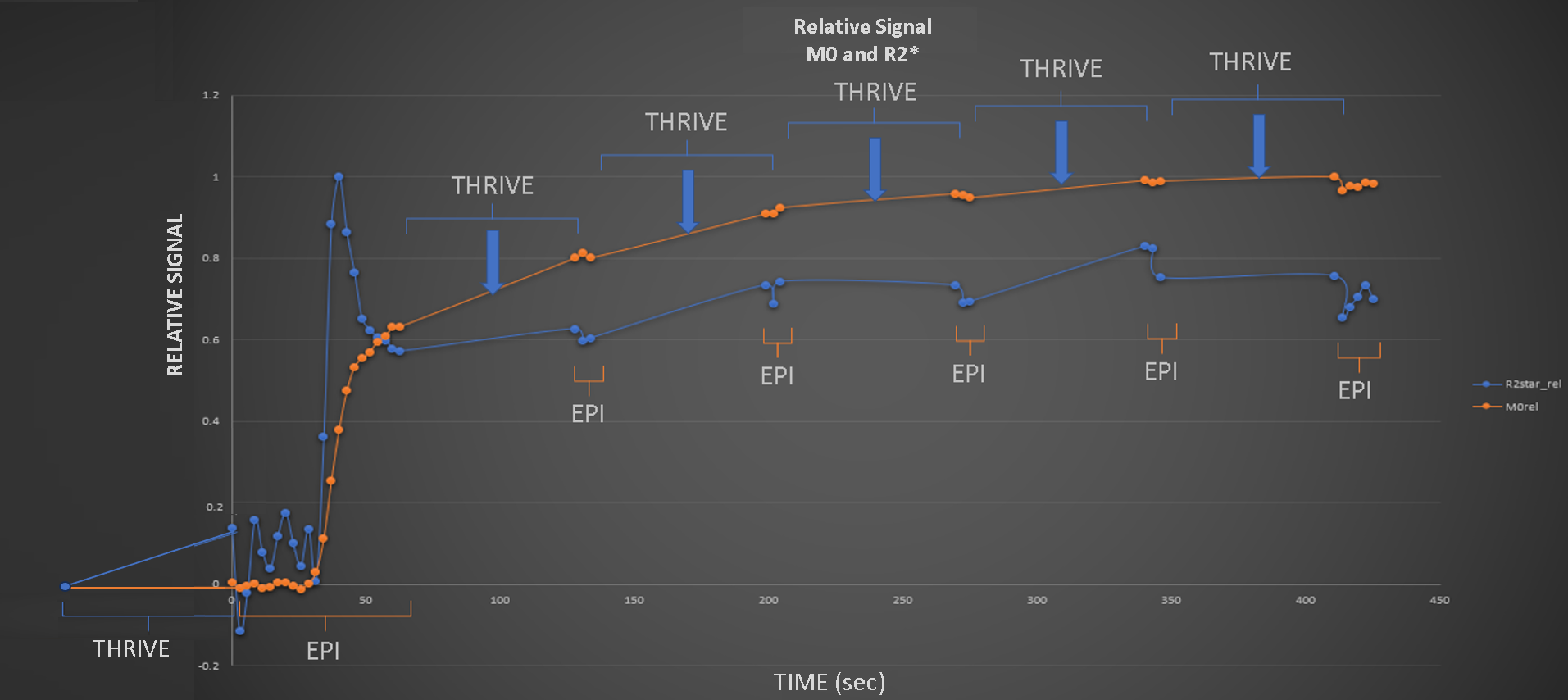
The MR pulse sequences utilized during the dynamic development of MR signals include the commonly employed T1-weighted THRIVE sequence, which is globally recognized for producing high spatial resolution images when a contrast agent is administered. By employing a multi-echo EPI pulse sequence, it becomes possible to visualize both the dynamic T1 signal (M0) and the dynamic R2* (R2*=1/T2*). During the scanning process, both the THRIVE and EPI sequences are applied in an alternating manner, resulting in two sets of dynamic MR data: one with high spatial information (THRIVE) and one with high temporal information (EPI).
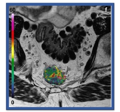
Observed rectal tumor heterogeneity. E. Grøvik et al, JMRI 2017.
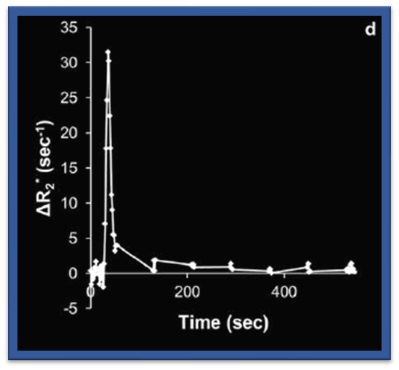
The dynamic R2* signal in rectal tumor. E. Grøvik et al, JMRI 2017.
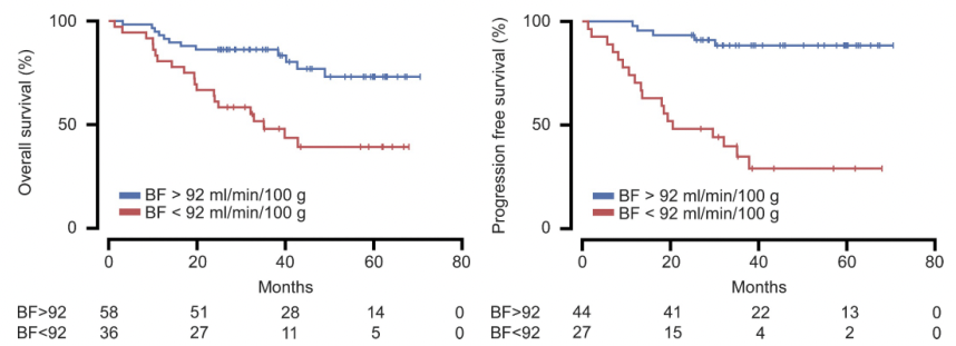
NordicCADs method predicts the evolution of rectal cancer for the next five years given standard treatment by measuring the blood flow (BF) in the cancer. K. Bakke et al, Radiology 2020.
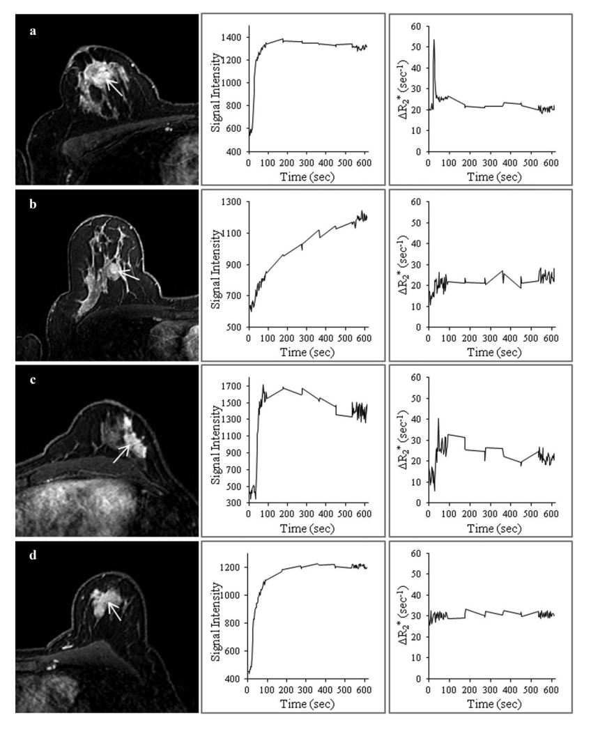
Signal Intensity curves (M0 and R2*) for different breast lesions. See Grøvik el al JMRI 2013 for details.
This study introduces the "split dynamic" method in MR imaging, combining high temporal and spatial resolution. It tested the accuracy and reliability of this method through simulations and patient data, focusing on kinetic parameter estimations. Results show effective evaluation of T1- and T2*-weighted characteristics, maintaining parameter reliability and adhering to spatial resolution standards.
Read MoreThis research focuses on applying the split dynamic MRI technique for assessing breast masses, combining high spatial resolution with dual-echo high temporal resolution data. The study involved 44 women, analyzing both BI-RADS interpretations and quantitative kinetic analysis. Findings indicate significant improvement in diagnostic accuracy for malignancy identification, highlighting the potential of split dynamic MRI as a more effective diagnostic tool in breast imaging.
Read MoreThis pilot study implements a dynamic contrast-based multi-echo MRI sequence in assessing rectal cancer, correlating histopathologic data with DCE- and DSC-MRI parameters. Data from 17 patients with resectable rectal cancer were analyzed. The study found significant associations between lower Ktrans and ΔR2* peak in the primary tumor and lymph node metastasis, demonstrating the feasibility and potential of this method in patient stratification for intensified multimodal treatment.
Read MoreThis research with 94 rectal cancer patients found that sex differences and tumor blood flow, measured using dynamic susceptibility contrast MRI, are key predictors of treatment response and survival. Lower blood flow was linked to worse treatment outcomes and shorter survival. Women typically had higher blood flow, impacting prognosis and treatment efficacy.
Read More